Must Read, Killing Cancer Cells Using Electric Potential, DMSO, Methylene Blue

OP
T

TreasureVibe

Member
Joined
Jul 3, 2016
Messages
1,941
Haidut adviced keto-acids for advanced cancer:

Glycine does contain nitrogen but inhibits iNOS and lowers NO. Arginine seems to directly promote tumor growth. I think in advanced cancer cases where all ingested protein is quickly turned into ammonia, the best approach would be to supplement with keto-acids as they would combine with the high ammonia cancer patients have and help synthesize protein. Not sure if you have heard of Peat's famous "potato protein soup" but that is the rationale behind it.
Cancer patients overwhelmingly have negative nitrogen balance due to elevated cortisol, and administering steroids like T, DHT or synthetic AAS like oxandrolone has been shown to reverse that negative balance so if potato protein soup is not an option administering these steroids can reverse the catabolic state and allow regular protein to be eaten.
Some links on arginine and cancer.
Conditionally Essential Amino Acids: Glycine And Arginine
Arginine Depletion May Be A Viable Approach For Cancer
Glycine May Treat Lung, Brain And Other Cancers
https://www.cell.com/cell-reports/pdf/S2211-1247(16)31799-5.pdf

Keto acid - Wikipedia

Keto acids or ketoacids (also called oxo acids or oxoacids) are organic compounds that contain a carboxylic acid group and a ketone group.[1] In several cases, the keto group is hydrated. The alpha-keto acids are especially important in biology as they are involved in the Krebs citric acid cycle and in glycolysis.[2]

Common types of keto acids include:

  • Alpha-keto acids, Alpha-ketoacids, or 2-oxoacids, such as pyruvic acid, have the keto group adjacent to the carboxylic acid. One important alpha-keto acid is oxaloacetic acid, a component of the Krebs cycle.[3] Another is alpha-ketoglutarate, is a 5-carbon ketoacid derived from glutamic acid. Alpha-ketoglutarate participates in cell signaling by functioning as a coenzyme,[4] and is commonly used in transamination reactions.
  • Beta-keto acids, Beta-ketoacids, or 3-oxoacids, such as acetoacetic acid, have the ketone group at the second carbon from the carboxylic acid. They can be formed by the Claisen condensation.
  • Gamma-keto acids, Gamma-ketoacids, or 4-oxoacids, such as levulinic acid, have the ketone group at the third carbon from the carboxylic acid.
Keto acids appear in a wide variety of anabolic pathways in metabolism, across living organisms. For instance, in plants (specifically, in hemlock, pitcher plants, and fool's parsley), 5-oxo-octanoic acid is converted in enzymatic and non-enzymatic steps into the cyclic class of coniine alkaloids.[citation needed]

When ingested sugars and carbohydrate levels are low, stored fats and proteins are the primary source of energy production. Glucogenic amino acids from proteins are converted to glucose. Ketogenic amino acids can be deaminated to produce alpha keto acids and ketone bodies.

Alpha keto acids are used primarily as energy for liver cells and in fatty acid synthesis, also in the liver.

Maybe either Oxaloacetic acid - Wikipedia or Acetoacetic acid - Wikipedia are good for cancer?

Acetoacetic acid (also diacetic acid) is the organic compound with the formula CH3COCH2COOH. It is the simplest beta-keto acid group, and like other members of this class, it is unstable. The methyl and ethyl esters, which are quite stable, are produced on a large scale industrially as precursors to dyes. Acetoacetic acid is a weak acid.[3]

Biochemistry
Under typical physiological conditions, acetoacetic acid exists as its conjugate base, acetoacetate.

Acetoacetate is produced in mitochondria of the liver from acetoacetyl Coenzyme A (CoA). First another acetyl group is added from acetyl CoA to form 3-hydroxy-3-methylgluteryl CoA, and then an acetyl CoA is lost from this yielding acetoacetate. The initial acetoacetate can come from the last cycle in the beta oxidation of a fatty acid, or it can be synthesized from two acetyl CoA molecules, catalyzed by thiolase.[4]

In mammals acetoacetate produced in the liver (along with the other two "ketone bodies") is released into the bloodstream as an energy source during periods of fasting, exercise, or as a result of type 1 diabetes mellitus.[5] First a CoA group is enzymatically transferred to it from succinyl CoA, converting it back to acetoacetyl CoA. This is then broken into two acetyl CoA molecules by thiolase, and these then enter the citric acid cycle. Heart muscle and renal cortex prefer acetoacetate over glucose. The brain uses acetoacetate when glucose levels are low due to fasting or diabetes.[6]

Acetoacetic acid - Wikipedia

Oxaloacetic acid (also known as oxalacetic acid) is a crystalline organic compound with the chemical formula HO2CC(O)CH2CO2H. Oxaloacetic acid, in the form of its conjugate base oxaloacetate, is a metabolic intermediate in many processes that occur in animals. It takes part in the gluconeogenesis, urea cycle, glyoxylate cycle, amino acid synthesis, fatty acid synthesis and citric acid cycle.[1]

Biosynthesis[edit]
Oxaloacetate forms in several ways in nature. A principal route is upon oxidation of L-malate, catalysed by malate dehydrogenase, in the citric acid cycle. Malate is also oxidized by succinate dehydrogenase in a slow reaction with the initial product being enol-oxaloacetate.[2]
It also arises from the condensation of pyruvate with carbonic acid, driven by the hydrolysis of ATP:

CH3C(O)CO2− + HCO3− + ATP → −O2CCH2C(O)CO2− + ADP + Pi
Occurring in the mesophyll of plants, this process proceeds via phosphoenolpyruvate, catalysed by pyruvate carboxylase.
Oxaloacetate can also arise from trans- or de- amination of aspartic acid.

Biochemical functions[edit]
Oxaloacetate is an intermediate of the citric acid cycle, where it reacts with acetyl-CoA to form citrate, catalysed by citrate synthase. It is also involved in gluconeogenesis, urea cycle, glyoxylate cycle, amino acid synthesis, and fatty acid synthesis. Oxaloacetate is also a potent inhibitor of Complex II.

Gluconeogenesis[edit]
Gluconeogenesis[1] is a metabolic pathway consisting of a series of eleven enzyme-catalyzed reactions, resulting in the generation of glucose from non-carbohydrates substrates. The beginning of this process takes place in the mitochondrial matrix, where pyruvate molecules are found. A pyruvate molecule is carboxylated by a pyruvate carboxylase enzyme, activated by a molecule each of ATP and water. This reaction results in the formation of oxaloacetate. NADH reduces oxaloacetate to malate. This transformation is needed to transport the molecule out of the mitochondria. Once in the cytosol, malate is oxidized to oxaloacetate again using NAD+. Then oxaloacetate remains in the cytosol, where the rest of reactions will take place. Oxaloacetate is later decarboxylated and phosphorylated by phosphoenolpyruvate carboxykinase and becomes 2-phosphoenolpyruvate using guanosine triphosphate (GTP) as phosphate source. Glucose is obtained after further downstream processing.

Urea cycle[edit]
The urea cycle is a metabolic pathway that results in the formation of urea using two ammonium molecules and one bicarbonate molecule.[1] This route commonly occurs in hepatocytes. The reactions related to the urea cycle produce NADH), and NADH can be produced in two different ways. One of these uses oxaloacetate. In the cytosol there are fumarate molecules. Fumarate can be transformed into malate by the actions of the enzyme fumarase. Malate is acted on by malate dehydrogenase to become oxaloacetate, producing a molecule of NADH. After that, oxaloacetate will be recycled to aspartate, as transaminases prefer these keto acids over the others. This recycling maintains the flow of nitrogen into the cell.


Glyoxylate cycle[edit]
The glyoxylate cycle is a variant of the citric acid cycle.[3] It is an anabolic pathway occurring in plants and bacteria utilizing the enzymes isocitrate lyase and malate synthase. Some intermediate steps of the cycle are slightly different from the citric acid cycle; nevertheless oxaloacetate has the same function in both processes.[1] This means that oxaloacetate in this cycle also acts as the primary reactant and final product. In fact the oxaloacetate is a net product of the glyoxylate cycle because its loop of the cycle incorporates two molecules of acetyl-CoA.

Fatty acid synthesis[edit]
In previous stages acetyl-CoA is transferred from the mitochondria to the cytoplasm where fatty acid synthase resides. The acetyl-CoA is transported as a citrate, which has been previously formed in the mitochondrial matrix from acetyl-coA and oxaloacetate. This reaction usually initiates the citric acid cycle, but when there is no need of energy it is transported to the cytoplasm where it is broken down to cytoplasmatic acetyl -CoA and oxaloacetate.

Another part of the cycle requires NADPH for the synthesis of fatty acids.[4] Part of this reducing power is generated when the cytosolic oxaloacetate is returned to the mitochondria as long as the internal mitochondrial layer is non-permeable for oxaloacetate. Firstly the oxaloacetate is reduced to malate using NADH. Then the malate is decarboxylated to pyruvate. Now this pyruvate can easily enter the mitochondria, where it is carboxylated again to oxaloacetate by pyruvate carboxylase. In this way, the transfer of acetyl-CoA that is from the mitochondria to the outside of the cell into the cytoplasm produces a molecule of NADH. The overall reaction, which is spontaneous, may be summarized as:

HCO3− + ATP + acetyl-CoA → ADP + Pi + malonyl-CoA
Amino acid synthesis[edit]
Six essential amino acids and three nonessential are synthesized from oxaloacetate and pyruvate.[5] Aspartate and alanine are formed from oxaloacetate and pyruvate, respectively, by transamination from glutamate. Asparagine, methionine, lysine and threonine are synthesized by aspartate, therefore given importance to oxaloacetate as without it, no aspartate would be formed and the following other amino acids would neither be produced.

Oxalate biosynthesis[edit]
Oxaloacetate produces oxalate by hydrolysis.[6]

oxaloacetate + H2O ⇌ oxalate + acetate
This process is catalyzed by the enzyme oxaloacetase.

Oxaloacetic acid - Wikipedia


Found this:

Acetoacetate is a metabolic inhibitor of cancer growth

Acetoacetate reduces growth and ATP concentration in cancer cell lines which over-express uncoupling protein 2. - PubMed - NCBI

Acetoacetate reduces growth and ATP concentration in cancer cell lines which over-express uncoupling protein 2

Addicted to AA (Acetoacetate): A Point of Convergence between Metabolism and BRAF Signaling

Prevention of Dietary-Fat-Fueled Ketogenesis Attenuates BRAF V600E Tumor Growth - ScienceDirect

Acetoacetate metabolism in AS-30D hepatoma cells

New Study Found Oxaloacetate Could Fight Cancer and Give Calorie Restriction Benefits

The metabolic fate of acetate in cancer

8) Oxaloacetate May Help Cancer
Glutamine is an important growth element for pancreatic cancer cells [R].

Studies have shown that reducing glutamine levels can stop cell growth of multiple forms of cancer [R].

Oxaloacetate has been shown to reduce glutamine levels and decrease cancer cell growth rates in mice [R].

It has also been shown to make chemotherapy and radiation treatment more effective [R].

Source: 15 Science-Based Health Benefits of Oxaloacetate - Selfhacked

Glutamine Addiction: A New Therapeutic Target in Cancer

Oxaloacetate induces apoptosis in HepG2 cells via inhibition of glycolysis

Oxaloacetate: 3 Outrageous Reasons This Molecule... - Nootropedia

Oxaloacetate: The Next Big Brain & Anti-Aging Supplement?

3 Ways an Oxaloacetate Supplement Can Protect You

CRONaxel is a US FDA registered combination of oxaloacetate and ascorbic acid for the clinical dietary management of the metabolic imbalances associated with certain cancers. (Oxaloacetate has the designation as an “orphan drug 12-3704” for the treatment of neuro-endocrine and glial tumors.)

Oxaloacetate is an obscure molecule that plays a role in energy production. Researchers have learned that it can also remove excess glutamate from the bloodstream. Glutamate can cause extensive brain damage in the event of a stroke or concussion, and it is a preferred fuel for many different types of cancer cells. Oxaloacetate’s removal of glutamate has a protective effect against these hazards.

Oxaloacetate increases energy production:
Cells can produce energy in two different ways. The more primitive, less effective method is glycolysis. The more advanced and efficient method is oxidative phosphorylation. This occurs in the mitochondria, and it produces eight times more energy than glycolysis—while using the same amount of fuel.[1] Oxaloacetate is an important part of the metabolic cycle that allows oxidative phosphorylation to occur. Without this molecule, cells are forced to rely more heavily on glycolysis.[2] This can lead to decreased production of energy and a decreased supply of blood sugar. In fact, studies indicate that a lack of oxaloacetate can cause energy production to fall by nearly 500%.[3]

Oxaloacetate starves cancer:
Cancerous cells rely on simple sugars and glutamate for their fuel supply, and this is especially true for brain tumors.[4] Glutamate also allows cancer cells to grow and divide.[5] When simple sugars and glutamate are abundant, cancer cells can produce more energy. This makes them more capable of spreading and more resistant to treatment. Fortunately, studies have shown that the consumption of an oxaloacetate supplement can decrease blood glutamate levels by 40%.[6,7] This leads to a reduction of tumor size and invasiveness.[8] Furthermore, patients who are given an oxaloacetate supplement experience an increased survival rate of 237%.[8]

Source with full text: CRONaxel Medical Food - Freeport Family Wellness Centre


Review Article | Published: 26 August 2016

Acetoacetate Assay Kit (Colorimetric) (ab180875) | Abcam
Acetoacetate (AcAc), a ß-ketoacid, is one of the three ketone bodies and is formed via condensation of two molecules of acetyl-CoA in liver mitochondria.


Conclusions
Our study suggests an important role of Glutamate oxaloacetate transaminase 1 (GOT1) to coordinate the glycolytic and the oxidative phosphorylation pathways in KRAS mutated cancer cells. GOT1 is crucial to provide oxaloacetate at low glucose levels, likely to maintain the redox homeostasis. Our data suggest GOT1 as a possible target in cancer therapy.

https://bmccancer.biomedcentral.com/articles/10.1186/s12885-018-4443-1


The metabolic fate of acetate in cancer
Abstract
Recent high-profile reports have reignited an interest in acetate metabolism in cancer. Acetyl-CoA synthetases that catalyse the conversion of acetate to acetyl-CoA have now been implicated in the growth of hepatocellular carcinoma, glioblastoma, breast cancer and prostate cancer. In this Review, we discuss how acetate functions as a nutritional source for tumours and as a regulator of cancer cell stress, and how preventing its (re)capture by cancer cells may provide an opportunity for therapeutic intervention.

Source: https://www.nature.com/articles/nrc.2016.87

Book source on cancer metabolism and oxaloacetate: Cancer

General information source: Is Cancer a Metabolic Disease? - ScienceDirect

Oxaloacetate is a new “Orphan Drug” for the treatment of Glioma

Study behind a paywall about CoA metabolism acetate and cancer: Acetate/acetyl-CoA metabolism associated with cancer fatty acid synthesis: Overview and application

Study: http://clincancerres.aacrjournals.o...rly/2015/03/26/1078-0432.CCR-14-1200.full.pdf

Acetate might be bad too in cancer:

The sources of acetyl-CoA and fatty acid synthesis
Acetyl-CoA is the precursor for the synthesis of fatty acids and cholesterol. Both glucose and glutamine can contribute to the generation of acetyl-CoA. Acetate has recently been shown to be yet another source of acetyl-CoA for many different cancer types, including breast, prostate, liver, primary glioblastomas and brain metastases. Some of these cancerous tissues incorporate acetate into fatty acids to support biomass production, whereas others have been shown to use acetate to fuel the TCA cycle (Comerford et al., 2014; Kamphorst et al., 2014; Mashimo et al., 2014; Schug et al., 2015). Mechanistically, acetate is ligated to CoA by the acyl-CoA synthetase short-chain family member 2 (ACSS2), an enzyme that is upregulated during conditions of metabolic stress such as low lipid availability and hypoxia. As such, acetate might become a crucial nutritional source in poorly vascularized regions of tumors.

Source: Cancer metabolism at a glance


Glyoxylic acid
Glyoxylic acid or oxoacetic acid is an organic compound. Together with acetic acid, glycolic acid, and oxalic acid, glyoxylic acid is one of the C2 carboxylic acids. It is a colourless solid that occurs naturally and is useful industrially.

"Historically glyoxylic acid was prepared from oxalic acid electrosynthetically:[7][8]
In organic synthesis, lead dioxide anodes were applied for the production of glyoxylic acid from oxalic acid in a sulfuric acid electrolyte.[9]

The conjugate base of glyoxylic acid is known as glyoxylate and is the form that the compound exists in solution at neutral pH. Glyoxylate is the byproduct of the amidation process in biosynthesis of several amidated peptides."

Source: Glyoxylic acid - Wikipedia


Glyoxylic... Hmm... Where have I heard that earlier... Glyoxylase?

I wonder how much of lactic acid produced is simply on account of the increased glyoxylase levels?

Glyoxylase I & II work in tandem to convert methylglyoxal into lactic acid, using glutathionine as a cofactor. Increased glyoxylase enzyme levels necessarily must lead to lower methylgloxal, lactic acid, and . . . perhaps even cancer:

Rulli, Antonio. "Expression of glyoxalase I and II in normal and breast cancer tissues." Breast cancer research and treatment (2001)

'A far higher activity level of glyoxylase I and II occurs in the tumor compared with pair-matched normal tissue, as shown by both spectrophotometrical assay and electrophoretic pattern. Such increased activities of glyoxylase I and II likely result from an enhanced enzyme synthesis as a consequence of increased expression of the respective genes in the tumoral tissue, as evidenced by northern blot.' ―Rulli


Paul Thornally is currently the leading authority on the methylglyoxal system, and has published many good articles on the topic. Doctor Thornally appears to have roughly 80 articles on methylglyoxal, but I do get the impression that he'd been given authorship credits in many of them simply on the basis of being advisor/editor of other peoples articles:

Thornalley, Paul J. "The glyoxalase system in health and disease." Molecular aspects of medicine (1993)

Other strong trends found in cancer cells are the increased expression of ornithine decarboxylase, increased stearoyl–CoA desaturase, and a diminished plasma membrane potential: Ornithine decarboxylase is of course the rate-limiting enzyme in polyamine synthesis, and polyamines physically bind dNA in a way that facilitates replication. Stearoyl–CoA desaturase tautologically desaturases stearate, when attached to coenzyme A, and increases membrane fluidity in this manner. For a cell to achieve one mitosis cycle, it needs to eventually double the amount of lipids on the cell membrane. You simply cannot achieve replication without the enzymes fatty acid synthase, stearoyl–CoA desaturase, and ornithine decarboxylase.

Lapachol and β-lapachone, found in pau d'arco, are two of the strongest glyoxylase inhibitors ever assayed. For inhibiting ornithine decarboxylase, thus decreasing polyamine synthesis, selenomethionine is a safe and natural amino acid to take—and one with other benefits besides: Some selenomethionine will eventually be metabolized, liberating inorganic selenium species that can be made available for selenocysteine synthesis. The enzyme glutathione peroxidase needs selenocysteine for reducing H₂O₂, and this enzyme can be synthesized using inorganic selenium (the inorganic selenium species is though to be added post-transcriptionally, meaning that ingesting selenocysteine proper is not required). By reducing cytosolic H₂O₂ concentrations, glutathione peroxidase lowers activation of NF-κB. This occurs because nuclear factor-κB is H₂O₂-sensitive, forming an internal disulfide bridge upon reacting with it. This achieves two ends: (1) it is then dissociated from its binding site, and (2) it is then compact enough to translocate into the cell's nucleus. The foregoing observations explain why inorganic selenium reduces cancer incidence, and also why selenomethionine—specifically—does that and more. [A person can also argue that it's the high methionine turnover of the prostate that accounts for selenomethionine's striking ability to reduce cancer there: the prostate synthesizes more polyamines than any other organ, and would of course need methionine to do so. This observation extends into the diagnostic realm, forming the basis for ¹¹C-methionine–PET imaging.]

CC: @Travis
 
Last edited:
OP
T

TreasureVibe

Member
Joined
Jul 3, 2016
Messages
1,941
In short,

There are studies that show that ketones can inhibit tumor growth and induce apoptosis in cancer cells.

A keto acid is a combination of a ketone with a carboxylic acid.

Glyoxylic acid is also named oxoacetic acid.

Acetate can be feeding tumor growth according to one source. Be aware of this. Please read this full source:

Cancer metabolism at a glance

"Acetate has recently been shown to be yet another source of acetyl-CoA for many different cancer types, including breast, prostate, liver, primary glioblastomas and brain metastases. Some of these cancerous tissues incorporate acetate into fatty acids to support biomass production, whereas others have been shown to use acetate to fuel the TCA cycle"

And please read this full source too:


"Recent high-profile reports have reignited an interest in acetate metabolism in cancer. Acetyl-CoA synthetases that catalyse the conversion of acetate to acetyl-CoA have now been implicated in the growth of hepatocellular carcinoma, glioblastoma, breast cancer and prostate cancer. In this Review, we discuss how acetate functions as a nutritional source for tumours and as a regulator of cancer cell stress, and how preventing its (re)capture by cancer cells may provide an opportunity for therapeutic intervention."

"Acetate can be used by tumour cells as an important bioenergetic fuel or as a nutritional source to support lipid biosynthesis."

Source: https://www.nature.com/articles/nrc.2016.87


Haidut recommends keto acids for advanced cancer, and there are studies that show that ketones can inhibit tumor growth and induce apoptosis in cancer cells.

There are also studies that show that a ketogenic diet improves outcome and life quality in cancer patients.

Ray Peat also was a proponent of a ketogenic diet earlier in his career.

There are keto acids that have acetate:

Acetoacetate and oxaloacetate for example.

There are studies about both showing they can inhibit tumor growth and induce apoptosis in cancer cells.

There is a new drug called CRONaxel which is FDA registered, which combines oxaloacetate with ascorbic acid for cancer:

CRONaxel is a US FDA registered combination of oxaloacetate and ascorbic acid for the clinical dietary management of the metabolic imbalances associated with certain cancers. (Oxaloacetate has the designation as an “orphan drug 12-3704” for the treatment of neuro-endocrine and glial tumors.)

Their source states the following:

Oxaloacetate is an obscure molecule that plays a role in energy production. Researchers have learned that it can also remove excess glutamate from the bloodstream. Glutamate can cause extensive brain damage in the event of a stroke or concussion, and it is a preferred fuel for many different types of cancer cells. Oxaloacetate’s removal of glutamate has a protective effect against these hazards.

Oxaloacetate increases energy production:
Cells can produce energy in two different ways. The more primitive, less effective method is glycolysis. The more advanced and efficient method is oxidative phosphorylation. This occurs in the mitochondria, and it produces eight times more energy than glycolysis—while using the same amount of fuel.[1] Oxaloacetate is an important part of the metabolic cycle that allows oxidative phosphorylation to occur. Without this molecule, cells are forced to rely more heavily on glycolysis.[2] This can lead to decreased production of energy and a decreased supply of blood sugar. In fact, studies indicate that a lack of oxaloacetate can cause energy production to fall by nearly 500%.[3]

Oxaloacetate starves cancer:
Cancerous cells rely on simple sugars and glutamate for their fuel supply, and this is especially true for brain tumors.[4] Glutamate also allows cancer cells to grow and divide.[5] When simple sugars and glutamate are abundant, cancer cells can produce more energy. This makes them more capable of spreading and more resistant to treatment. Fortunately, studies have shown that the consumption of an oxaloacetate supplement can decrease blood glutamate levels by 40%.[6,7] This leads to a reduction of tumor size and invasiveness.[8] Furthermore, patients who are given an oxaloacetate supplement experience an increased survival rate of 237%.[8]


Acetoacetate still remains interesting too for treating cancer.

Acetoacetate is (also) made in the liver:
Acetoacetate Assay Kit (Colorimetric) (ab180875) | Abcam
Acetoacetate (AcAc), a ß-ketoacid, is one of the three ketone bodies and is formed via condensation of two molecules of acetyl-CoA in liver mitochondria.

CC @Travis @Obi-wan

Sources on CRONaxel:
CRONaxel Medical Food - Freeport Family Wellness Centre
Article with some details about the product that raise skepticism CRONaxal - RationalWiki
Product with similar ingredients https://www.amazon.com/Fractal-Health-Oxaloacetate-OAA/dp/B01870220A#customerReviews
 
Last edited:
OP
T

TreasureVibe

Member
Joined
Jul 3, 2016
Messages
1,941
This is also very interesting, from the first CRONaxel source under "Treatments" > "Metabolic Cancer Control":

Master Amino Acid Pattern (MAP)


The main issue with putting a person living with cancer on a high-protein, low carbohydrate diet to induce ketosis and ‘starve’ the cancer of its source of sugar fuels, is that a high-protein diet puts a great strain on the liver and kidneys of anyone doing the high-protein ketogenic diet and especially stresses people weakened from cancer and toxic treatments for cancer (chemotherapy). This happens because the high-protein diet produces high levels of metabolic waste and even some sugar, so it doesn’t ‘starve’ the cancer the way hundreds of websites seem to suggest.

The good news is that there is a different (and completely safe) diet that induces ketosis without producing metabolic waste products that stress the liver and kidneys. A diet that is high in essential, healthy fatty acids (I hope they don't mean PUFA with this) and restricted in protein and carbohydrates in theory would produce the desired ketones and ‘starve’ the cancer of fuel from carbohydrate and protein conversion to glucose and other sugars.

The Master Amino Acid Pattern (MAP) protects the body from being depleted of essential amino acids while inducing ketone production. The interesting and unique features of MAP are that the “Net Nitrogen Utilization for Body Protein Synthesis” is 99% while dietary protein NNU is only 16% to 32% depending on the protein source. The MAP also absorbs in the body in 23 minutes compared to 2-6 hours for other proteins. Taking MAP while on a high fatty acid diet ketogenic diet will maintain a safe healthy nitrogen balance.

Source: Master Amino Acid Pattern - Freeport Family Wellness Centre
Source on what they possibly mean with essential healthy fatty acids (where they include coconut oil): Medium Chain Triglycerides - Freeport Family Wellness Centre
More on MAP: FAQs

Btw on methylene blue:

Here's a mice study showing it lowered natural killer T-cells: Methylene Blue Selectively Inhibits B Cell Development and E2A Activity

"Results MB decreased the proliferation of precursor B cell lines, REH, NALM-6 and BAF3-p185 cells compared to myeloid cell lines, CCRF and CMK. Similar effects of MB were also seen with normal B and myeloid progenitors as assayed by methylcellulose colony formation assays with murine bone marrow cells. Similarly, there is a significant decrease in peripheral B cells as compared to granulocytes, macrophages, T cells and NK cells when the mice were treated with 1000 μM of methylene blue in their drinking water as compared to those that remained untreated. MB caused apoptosis and decreased the S phase fraction of treated NALM-6 cells. In addition, we found that MB inhibited E2A activity using an E2A-specific luciferase reporter assay."

I don't really know how to interpret what they've wrote here so it might say something else as well..
 
Last edited:

Obi-wan

Member
Joined
Mar 16, 2017
Messages
1,120
upload_2018-7-4_10-3-41.png


Adrenal hormone pathways. All lines leading to Estrone and Estradiol are aromatase lines. 90% of hormones are activated by the pituitary gland. 10% by the adrenal glands.
 

Obi-wan

Member
Joined
Mar 16, 2017
Messages
1,120
Haidut adviced keto-acids for advanced cancer:


Keto acid - Wikipedia

Keto acids or ketoacids (also called oxo acids or oxoacids) are organic compounds that contain a carboxylic acid group and a ketone group.[1] In several cases, the keto group is hydrated. The alpha-keto acids are especially important in biology as they are involved in the Krebs citric acid cycle and in glycolysis.[2]

Common types of keto acids include:

  • Alpha-keto acids, Alpha-ketoacids, or 2-oxoacids, such as pyruvic acid, have the keto group adjacent to the carboxylic acid. One important alpha-keto acid is oxaloacetic acid, a component of the Krebs cycle.[3] Another is alpha-ketoglutarate, is a 5-carbon ketoacid derived from glutamic acid. Alpha-ketoglutarate participates in cell signaling by functioning as a coenzyme,[4] and is commonly used in transamination reactions.
  • Beta-keto acids, Beta-ketoacids, or 3-oxoacids, such as acetoacetic acid, have the ketone group at the second carbon from the carboxylic acid. They can be formed by the Claisen condensation.
  • Gamma-keto acids, Gamma-ketoacids, or 4-oxoacids, such as levulinic acid, have the ketone group at the third carbon from the carboxylic acid.
Keto acids appear in a wide variety of anabolic pathways in metabolism, across living organisms. For instance, in plants (specifically, in hemlock, pitcher plants, and fool's parsley), 5-oxo-octanoic acid is converted in enzymatic and non-enzymatic steps into the cyclic class of coniine alkaloids.[citation needed]

When ingested sugars and carbohydrate levels are low, stored fats and proteins are the primary source of energy production. Glucogenic amino acids from proteins are converted to glucose. Ketogenic amino acids can be deaminated to produce alpha keto acids and ketone bodies.

Alpha keto acids are used primarily as energy for liver cells and in fatty acid synthesis, also in the liver.

Maybe either Oxaloacetic acid - Wikipedia or Acetoacetic acid - Wikipedia are good for cancer?

Acetoacetic acid (also diacetic acid) is the organic compound with the formula CH3COCH2COOH. It is the simplest beta-keto acid group, and like other members of this class, it is unstable. The methyl and ethyl esters, which are quite stable, are produced on a large scale industrially as precursors to dyes. Acetoacetic acid is a weak acid.[3]

Biochemistry
Under typical physiological conditions, acetoacetic acid exists as its conjugate base, acetoacetate.

Acetoacetate is produced in mitochondria of the liver from acetoacetyl Coenzyme A (CoA). First another acetyl group is added from acetyl CoA to form 3-hydroxy-3-methylgluteryl CoA, and then an acetyl CoA is lost from this yielding acetoacetate. The initial acetoacetate can come from the last cycle in the beta oxidation of a fatty acid, or it can be synthesized from two acetyl CoA molecules, catalyzed by thiolase.[4]

In mammals acetoacetate produced in the liver (along with the other two "ketone bodies") is released into the bloodstream as an energy source during periods of fasting, exercise, or as a result of type 1 diabetes mellitus.[5] First a CoA group is enzymatically transferred to it from succinyl CoA, converting it back to acetoacetyl CoA. This is then broken into two acetyl CoA molecules by thiolase, and these then enter the citric acid cycle. Heart muscle and renal cortex prefer acetoacetate over glucose. The brain uses acetoacetate when glucose levels are low due to fasting or diabetes.[6]

Acetoacetic acid - Wikipedia

Oxaloacetic acid (also known as oxalacetic acid) is a crystalline organic compound with the chemical formula HO2CC(O)CH2CO2H. Oxaloacetic acid, in the form of its conjugate base oxaloacetate, is a metabolic intermediate in many processes that occur in animals. It takes part in the gluconeogenesis, urea cycle, glyoxylate cycle, amino acid synthesis, fatty acid synthesis and citric acid cycle.[1]

Biosynthesis[edit]
Oxaloacetate forms in several ways in nature. A principal route is upon oxidation of L-malate, catalysed by malate dehydrogenase, in the citric acid cycle. Malate is also oxidized by succinate dehydrogenase in a slow reaction with the initial product being enol-oxaloacetate.[2]
It also arises from the condensation of pyruvate with carbonic acid, driven by the hydrolysis of ATP:

CH3C(O)CO2− + HCO3− + ATP → −O2CCH2C(O)CO2− + ADP + Pi
Occurring in the mesophyll of plants, this process proceeds via phosphoenolpyruvate, catalysed by pyruvate carboxylase.
Oxaloacetate can also arise from trans- or de- amination of aspartic acid.

Biochemical functions[edit]
Oxaloacetate is an intermediate of the citric acid cycle, where it reacts with acetyl-CoA to form citrate, catalysed by citrate synthase. It is also involved in gluconeogenesis, urea cycle, glyoxylate cycle, amino acid synthesis, and fatty acid synthesis. Oxaloacetate is also a potent inhibitor of Complex II.

Gluconeogenesis[edit]
Gluconeogenesis[1] is a metabolic pathway consisting of a series of eleven enzyme-catalyzed reactions, resulting in the generation of glucose from non-carbohydrates substrates. The beginning of this process takes place in the mitochondrial matrix, where pyruvate molecules are found. A pyruvate molecule is carboxylated by a pyruvate carboxylase enzyme, activated by a molecule each of ATP and water. This reaction results in the formation of oxaloacetate. NADH reduces oxaloacetate to malate. This transformation is needed to transport the molecule out of the mitochondria. Once in the cytosol, malate is oxidized to oxaloacetate again using NAD+. Then oxaloacetate remains in the cytosol, where the rest of reactions will take place. Oxaloacetate is later decarboxylated and phosphorylated by phosphoenolpyruvate carboxykinase and becomes 2-phosphoenolpyruvate using guanosine triphosphate (GTP) as phosphate source. Glucose is obtained after further downstream processing.

Urea cycle[edit]
The urea cycle is a metabolic pathway that results in the formation of urea using two ammonium molecules and one bicarbonate molecule.[1] This route commonly occurs in hepatocytes. The reactions related to the urea cycle produce NADH), and NADH can be produced in two different ways. One of these uses oxaloacetate. In the cytosol there are fumarate molecules. Fumarate can be transformed into malate by the actions of the enzyme fumarase. Malate is acted on by malate dehydrogenase to become oxaloacetate, producing a molecule of NADH. After that, oxaloacetate will be recycled to aspartate, as transaminases prefer these keto acids over the others. This recycling maintains the flow of nitrogen into the cell.


Glyoxylate cycle[edit]
The glyoxylate cycle is a variant of the citric acid cycle.[3] It is an anabolic pathway occurring in plants and bacteria utilizing the enzymes isocitrate lyase and malate synthase. Some intermediate steps of the cycle are slightly different from the citric acid cycle; nevertheless oxaloacetate has the same function in both processes.[1] This means that oxaloacetate in this cycle also acts as the primary reactant and final product. In fact the oxaloacetate is a net product of the glyoxylate cycle because its loop of the cycle incorporates two molecules of acetyl-CoA.

Fatty acid synthesis[edit]
In previous stages acetyl-CoA is transferred from the mitochondria to the cytoplasm where fatty acid synthase resides. The acetyl-CoA is transported as a citrate, which has been previously formed in the mitochondrial matrix from acetyl-coA and oxaloacetate. This reaction usually initiates the citric acid cycle, but when there is no need of energy it is transported to the cytoplasm where it is broken down to cytoplasmatic acetyl -CoA and oxaloacetate.

Another part of the cycle requires NADPH for the synthesis of fatty acids.[4] Part of this reducing power is generated when the cytosolic oxaloacetate is returned to the mitochondria as long as the internal mitochondrial layer is non-permeable for oxaloacetate. Firstly the oxaloacetate is reduced to malate using NADH. Then the malate is decarboxylated to pyruvate. Now this pyruvate can easily enter the mitochondria, where it is carboxylated again to oxaloacetate by pyruvate carboxylase. In this way, the transfer of acetyl-CoA that is from the mitochondria to the outside of the cell into the cytoplasm produces a molecule of NADH. The overall reaction, which is spontaneous, may be summarized as:

HCO3− + ATP + acetyl-CoA → ADP + Pi + malonyl-CoA
Amino acid synthesis[edit]
Six essential amino acids and three nonessential are synthesized from oxaloacetate and pyruvate.[5] Aspartate and alanine are formed from oxaloacetate and pyruvate, respectively, by transamination from glutamate. Asparagine, methionine, lysine and threonine are synthesized by aspartate, therefore given importance to oxaloacetate as without it, no aspartate would be formed and the following other amino acids would neither be produced.

Oxalate biosynthesis[edit]
Oxaloacetate produces oxalate by hydrolysis.[6]

oxaloacetate + H2O ⇌ oxalate + acetate
This process is catalyzed by the enzyme oxaloacetase.

Oxaloacetic acid - Wikipedia


Found this:

Acetoacetate is a metabolic inhibitor of cancer growth

Acetoacetate reduces growth and ATP concentration in cancer cell lines which over-express uncoupling protein 2. - PubMed - NCBI

Acetoacetate reduces growth and ATP concentration in cancer cell lines which over-express uncoupling protein 2

Addicted to AA (Acetoacetate): A Point of Convergence between Metabolism and BRAF Signaling

Prevention of Dietary-Fat-Fueled Ketogenesis Attenuates BRAF V600E Tumor Growth - ScienceDirect

Acetoacetate metabolism in AS-30D hepatoma cells

New Study Found Oxaloacetate Could Fight Cancer and Give Calorie Restriction Benefits

The metabolic fate of acetate in cancer

8) Oxaloacetate May Help Cancer
Glutamine is an important growth element for pancreatic cancer cells [R].

Studies have shown that reducing glutamine levels can stop cell growth of multiple forms of cancer [R].

Oxaloacetate has been shown to reduce glutamine levels and decrease cancer cell growth rates in mice [R].

It has also been shown to make chemotherapy and radiation treatment more effective [R].

Source: 15 Science-Based Health Benefits of Oxaloacetate - Selfhacked

Glutamine Addiction: A New Therapeutic Target in Cancer

Oxaloacetate induces apoptosis in HepG2 cells via inhibition of glycolysis

Oxaloacetate: 3 Outrageous Reasons This Molecule... - Nootropedia

Oxaloacetate: The Next Big Brain & Anti-Aging Supplement?

3 Ways an Oxaloacetate Supplement Can Protect You

CRONaxel is a US FDA registered combination of oxaloacetate and ascorbic acid for the clinical dietary management of the metabolic imbalances associated with certain cancers. (Oxaloacetate has the designation as an “orphan drug 12-3704” for the treatment of neuro-endocrine and glial tumors.)

Oxaloacetate is an obscure molecule that plays a role in energy production. Researchers have learned that it can also remove excess glutamate from the bloodstream. Glutamate can cause extensive brain damage in the event of a stroke or concussion, and it is a preferred fuel for many different types of cancer cells. Oxaloacetate’s removal of glutamate has a protective effect against these hazards.

Oxaloacetate increases energy production:
Cells can produce energy in two different ways. The more primitive, less effective method is glycolysis. The more advanced and efficient method is oxidative phosphorylation. This occurs in the mitochondria, and it produces eight times more energy than glycolysis—while using the same amount of fuel.[1] Oxaloacetate is an important part of the metabolic cycle that allows oxidative phosphorylation to occur. Without this molecule, cells are forced to rely more heavily on glycolysis.[2] This can lead to decreased production of energy and a decreased supply of blood sugar. In fact, studies indicate that a lack of oxaloacetate can cause energy production to fall by nearly 500%.[3]

Oxaloacetate starves cancer:
Cancerous cells rely on simple sugars and glutamate for their fuel supply, and this is especially true for brain tumors.[4] Glutamate also allows cancer cells to grow and divide.[5] When simple sugars and glutamate are abundant, cancer cells can produce more energy. This makes them more capable of spreading and more resistant to treatment. Fortunately, studies have shown that the consumption of an oxaloacetate supplement can decrease blood glutamate levels by 40%.[6,7] This leads to a reduction of tumor size and invasiveness.[8] Furthermore, patients who are given an oxaloacetate supplement experience an increased survival rate of 237%.[8]

Source with full text: CRONaxel Medical Food - Freeport Family Wellness Centre


Review Article | Published: 26 August 2016

Acetoacetate Assay Kit (Colorimetric) (ab180875) | Abcam
Acetoacetate (AcAc), a ß-ketoacid, is one of the three ketone bodies and is formed via condensation of two molecules of acetyl-CoA in liver mitochondria.


Conclusions
Our study suggests an important role of Glutamate oxaloacetate transaminase 1 (GOT1) to coordinate the glycolytic and the oxidative phosphorylation pathways in KRAS mutated cancer cells. GOT1 is crucial to provide oxaloacetate at low glucose levels, likely to maintain the redox homeostasis. Our data suggest GOT1 as a possible target in cancer therapy.

https://bmccancer.biomedcentral.com/articles/10.1186/s12885-018-4443-1


The metabolic fate of acetate in cancer
Abstract
Recent high-profile reports have reignited an interest in acetate metabolism in cancer. Acetyl-CoA synthetases that catalyse the conversion of acetate to acetyl-CoA have now been implicated in the growth of hepatocellular carcinoma, glioblastoma, breast cancer and prostate cancer. In this Review, we discuss how acetate functions as a nutritional source for tumours and as a regulator of cancer cell stress, and how preventing its (re)capture by cancer cells may provide an opportunity for therapeutic intervention.

Source: https://www.nature.com/articles/nrc.2016.87

Book source on cancer metabolism and oxaloacetate: Cancer

General information source: Is Cancer a Metabolic Disease? - ScienceDirect

Oxaloacetate is a new “Orphan Drug” for the treatment of Glioma

Study behind a paywall about CoA metabolism acetate and cancer: Acetate/acetyl-CoA metabolism associated with cancer fatty acid synthesis: Overview and application

Study: http://clincancerres.aacrjournals.o...rly/2015/03/26/1078-0432.CCR-14-1200.full.pdf

Acetate might be bad too in cancer:

The sources of acetyl-CoA and fatty acid synthesis
Acetyl-CoA is the precursor for the synthesis of fatty acids and cholesterol. Both glucose and glutamine can contribute to the generation of acetyl-CoA. Acetate has recently been shown to be yet another source of acetyl-CoA for many different cancer types, including breast, prostate, liver, primary glioblastomas and brain metastases. Some of these cancerous tissues incorporate acetate into fatty acids to support biomass production, whereas others have been shown to use acetate to fuel the TCA cycle (Comerford et al., 2014; Kamphorst et al., 2014; Mashimo et al., 2014; Schug et al., 2015). Mechanistically, acetate is ligated to CoA by the acyl-CoA synthetase short-chain family member 2 (ACSS2), an enzyme that is upregulated during conditions of metabolic stress such as low lipid availability and hypoxia. As such, acetate might become a crucial nutritional source in poorly vascularized regions of tumors.

Source: Cancer metabolism at a glance


Glyoxylic acid
Glyoxylic acid or oxoacetic acid is an organic compound. Together with acetic acid, glycolic acid, and oxalic acid, glyoxylic acid is one of the C2 carboxylic acids. It is a colourless solid that occurs naturally and is useful industrially.

"Historically glyoxylic acid was prepared from oxalic acid electrosynthetically:[7][8]
In organic synthesis, lead dioxide anodes were applied for the production of glyoxylic acid from oxalic acid in a sulfuric acid electrolyte.[9]

The conjugate base of glyoxylic acid is known as glyoxylate and is the form that the compound exists in solution at neutral pH. Glyoxylate is the byproduct of the amidation process in biosynthesis of several amidated peptides."

Source: Glyoxylic acid - Wikipedia


Glyoxylic... Hmm... Where have I heard that earlier... Glyoxylase?


CC: @Travis


There are a lot of pathways to metabolism (all use acetyl-CoA) and a lot of "if's" here. Many FAS inhibitors are mentioned on this forum, Aspirin, Niacinamide, etc...

Plus many on this forum (including me) practice low PUFA consumption (linoleic acid) and higher stearic acid consumption
 
Last edited:
OP
T

TreasureVibe

Member
Joined
Jul 3, 2016
Messages
1,941
There are a lot of pathways to metabolism (all use acetyl-CoA) and a lot of "if's" here. Many FAS inhibitors are mentioned on this forum, Aspirin, Niacinamide, etc...

Plus many on this forum (including me) practice low PUFA consumption (linoleic acid) and higher stearic acid consumption


What do you think of this though?
In short,

There are studies that show that ketones can inhibit tumor growth and induce apoptosis in cancer cells.

A keto acid is a combination of a ketone with a carboxylic acid.

Glyoxylic acid is also named oxoacetic acid.

Acetate can be feeding tumor growth according to one source. Be aware of this. Please read this full source:

Cancer metabolism at a glance

"Acetate has recently been shown to be yet another source of acetyl-CoA for many different cancer types, including breast, prostate, liver, primary glioblastomas and brain metastases. Some of these cancerous tissues incorporate acetate into fatty acids to support biomass production, whereas others have been shown to use acetate to fuel the TCA cycle"

And please read this full source too:


"Recent high-profile reports have reignited an interest in acetate metabolism in cancer. Acetyl-CoA synthetases that catalyse the conversion of acetate to acetyl-CoA have now been implicated in the growth of hepatocellular carcinoma, glioblastoma, breast cancer and prostate cancer. In this Review, we discuss how acetate functions as a nutritional source for tumours and as a regulator of cancer cell stress, and how preventing its (re)capture by cancer cells may provide an opportunity for therapeutic intervention."

"Acetate can be used by tumour cells as an important bioenergetic fuel or as a nutritional source to support lipid biosynthesis."

Source: https://www.nature.com/articles/nrc.2016.87


And this one?
There is a new drug called CRONaxel which is FDA registered, which combines oxaloacetate with ascorbic acid for cancer:

CRONaxel is a US FDA registered combination of oxaloacetate and ascorbic acid for the clinical dietary management of the metabolic imbalances associated with certain cancers. (Oxaloacetate has the designation as an “orphan drug 12-3704” for the treatment of neuro-endocrine and glial tumors.)

Their source states the following:

Oxaloacetate is an obscure molecule that plays a role in energy production. Researchers have learned that it can also remove excess glutamate from the bloodstream. Glutamate can cause extensive brain damage in the event of a stroke or concussion, and it is a preferred fuel for many different types of cancer cells. Oxaloacetate’s removal of glutamate has a protective effect against these hazards.

Oxaloacetate increases energy production:
Cells can produce energy in two different ways. The more primitive, less effective method is glycolysis. The more advanced and efficient method is oxidative phosphorylation. This occurs in the mitochondria, and it produces eight times more energy than glycolysis—while using the same amount of fuel.[1] Oxaloacetate is an important part of the metabolic cycle that allows oxidative phosphorylation to occur. Without this molecule, cells are forced to rely more heavily on glycolysis.[2] This can lead to decreased production of energy and a decreased supply of blood sugar. In fact, studies indicate that a lack of oxaloacetate can cause energy production to fall by nearly 500%.[3]

Oxaloacetate starves cancer:
Cancerous cells rely on simple sugars and glutamate for their fuel supply, and this is especially true for brain tumors.[4] Glutamate also allows cancer cells to grow and divide.[5] When simple sugars and glutamate are abundant, cancer cells can produce more energy. This makes them more capable of spreading and more resistant to treatment. Fortunately, studies have shown that the consumption of an oxaloacetate supplement can decrease blood glutamate levels by 40%.[6,7] This leads to a reduction of tumor size and invasiveness.[8] Furthermore, patients who are given an oxaloacetate supplement experience an increased survival rate of 237%.[8]


Acetoacetate still remains interesting too for treating cancer.

Acetoacetate is (also) made in the liver:
Acetoacetate Assay Kit (Colorimetric) (ab180875) | Abcam
Acetoacetate (AcAc), a ß-ketoacid, is one of the three ketone bodies and is formed via condensation of two molecules of acetyl-CoA in liver mitochondria.

CC @Travis @Obi-wan

Sources on CRONaxel:
CRONaxel Medical Food - Freeport Family Wellness Centre
Article with some details about the product that raise skepticism CRONaxal - RationalWiki
Product with similar ingredients https://www.amazon.com/Fractal-Health-Oxaloacetate-OAA/dp/B01870220A#customerReviews
 
Last edited:

Obi-wan

Member
Joined
Mar 16, 2017
Messages
1,120


Interesting flow chart by Wikipedia

@Travis, want to comment?
 
OP
T

TreasureVibe

Member
Joined
Jul 3, 2016
Messages
1,941
"Recent high-profile reports have reignited an interest in acetate metabolism in cancer. Acetyl-CoA synthetases that catalyse the conversion of acetate to acetyl-CoA have now been implicated in the growth of hepatocellular carcinoma, glioblastoma, breast cancer and prostate cancer. In this Review, we discuss how acetate functions as a nutritional source for tumours and as a regulator of cancer cell stress, and how preventing its (re)capture by cancer cells may provide an opportunity for therapeutic intervention."

I really hope that this study is biased...
 

Obi-wan

Member
Joined
Mar 16, 2017
Messages
1,120
IMO, Intracellular Ph of a cancer cell is alkaline due to sodium, calcium and bulk water. Extra cellular Ph is acidic due to high Lactic acid build up due to high Pyruvate buildup. We talked about apple cider vinager and baking soda which when combined produces sodium and potassium acetate and carbon dioxide. The carbon dioxide will remove the Lactic acid, the potassium acetate will create a proper electric potential (Ted from Bangkok) and converts to acetyl-CoA intracellular which operates the citric acid cycle and produces ATP and more carbon dioxide. This creates proper cellular metabolism and fixes the defective metabolism of a cancer cell over time. I have been experimenting with ACV/BS using Ph paper on urine to determine the amount of BS (now 1/4 teaspoon with 2 tablespoons of ACV). I have also been experimenting with frequency (now 1 to 2 times per day) Keeping this in mind "Pyruvate dehydrogenase is inhibited when one or more of the three following ratios are increased:
ATP/ADP, NADH/NAD+ and acetyl-CoA/CoA.
 
Last edited:

Obi-wan

Member
Joined
Mar 16, 2017
Messages
1,120
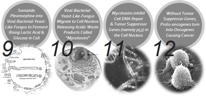
But pathogens must be involved, because they block the tumor suppressing genes that are supposed to induce apoptosis, supposedly.

See:

300437_orig.png


Check this website and press the white button in the upper left corner for more information:
PSYCHO-ONCOLOGY: HOW CHRONIC STRESS CAUSES CANCER OVER 6 PHASES
Also look at the schematic that's made, showing the steps of how cancer presumably develops.
Not condoning that website's information but it can come in handy.

Btw check this out on ketone supplementation:
Ketone supplementation decreases tumor cell viability and prolongs survival of mice with metastatic cancer
How I Used Ketone Bodies to Help Reverse My Brain Cancer | The Truth About Cancer
How the Ketogenic Diet Weakens Cancer Cells
Tumor Cells Growth and Survival Time with the Ketogenic Diet in Animal Models: A Systematic Review

However there are sceptics and it might be bad in some cancers: Ketogenic Diet For Cancer? Dr. Gonzalez Dismantles The Diet

Not sure if it works or is helpful, but it sure is interesting.


IMO, this is EXACTLY what happens on a biological level. But change the terrain to acidic from alkaline and the somatid is no longer pathogenic...
 
OP
T

TreasureVibe

Member
Joined
Jul 3, 2016
Messages
1,941
IMO, Intracellular Ph of a cancer cell is alkaline due to sodium, calcium and bulk water. Extra cellular Ph is acidic due to high Lactic acid build up due to high Pyruvate buildup. We talked about apple cider vinager and baking soda which when combined produces sodium and potassium acetate and carbon dioxide. The carbon dioxide will remove the Lactic acid, the potassium acetate will create a proper electric potential (Ted from Bangkok) and converts to acetyl co-enzyme intracellular which operates the citric acid cycle and produces ATP and more carbon dioxide. This creates proper cellular metabolism and fixes the defective metabolism of a cancer cell over time. I have been experimenting with ACV/BS using Ph paper on urine to determine the amount of BS (now 1/4 teaspoon with 2 tablespoons of ACV). I have also been experimenting with frequency (now 1 to 2 times per day) Keeping this in mind "Pyruvate dehydrogenase is inhibited when one or more of the three following ratios are increased:
ATP/ADP, NADH/NAD+ and acetyl-CoA/CoA.
A great summary and with the addition of your own experiences it pretty much seals it.. But, if acetate creates a proper electric potential, why wouldn't another carboxylic acid from the Krebs cycle do the same, like citrate? And by supplementing acetate, wouldn't a different carboxylic acid in the Krebs cycle become deficient? Why exactly is citric acid/citrate bad according to Dr. Peat, because it can cause dormant cancer cells to wake, yet acetate is perfectly fine? Wouldn't supplementation of all Krebs cycle nutrients be smart to make sure you're not deficient in any of them?

In other words, what makes acetate so special?
 
OP
T

TreasureVibe

Member
Joined
Jul 3, 2016
Messages
1,941
IMO, this is EXACTLY what happens on a biological level. But change the terrain to acidic from alkaline and the somatid is no longer pathogenic...
Its not the somatid that travels to the nucleus and releases mycotoxins, those are ACTUAL pathogens.
 
B

Braveheart

Guest
IMO, Intracellular Ph of a cancer cell is alkaline due to sodium, calcium and bulk water. Extra cellular Ph is acidic due to high Lactic acid build up due to high Pyruvate buildup. We talked about apple cider vinager and baking soda which when combined produces sodium and potassium acetate and carbon dioxide. The carbon dioxide will remove the Lactic acid, the potassium acetate will create a proper electric potential (Ted from Bangkok) and converts to acetyl co-enzyme intracellular which operates the citric acid cycle and produces ATP and more carbon dioxide. This creates proper cellular metabolism and fixes the defective metabolism of a cancer cell over time. I have been experimenting with ACV/BS using Ph paper on urine to determine the amount of BS (now 1/4 teaspoon with 2 tablespoons of ACV). I have also been experimenting with frequency (now 1 to 2 times per day) Keeping this in mind "Pyruvate dehydrogenase is inhibited when one or more of the three following ratios are increased:
ATP/ADP, NADH/NAD+ and acetyl-CoA/CoA.
You're on fire Obi-mon!...I settled on 1/4 tsp BS w 2 tbsp ACV...it felt right...couldn't find ph strips...1 or 2 times per day....still plan on going the lime route soon.
 

Obi-wan

Member
Joined
Mar 16, 2017
Messages
1,120
You're on fire Obi-mon!...I settled on 1/4 tsp BS w 2 tbsp ACV...it felt right...couldn't find ph strips...1 or 2 times per day....still plan on going the lime route soon.
I can’t do the mix at night because it keeps me awake but I will do BS (1/4 teaspoon) and water (8 ounces) at night if I think I will get heartburn...morning urine PH has been around 6.5-6.8 (dark green) used to be 5.5 (pale yellow). Body getting rid of high Lactic acid at 5.5...
 
Last edited:

Obi-wan

Member
Joined
Mar 16, 2017
Messages
1,120
A great summary and with the addition of your own experiences it pretty much seals it.. But, if acetate creates a proper electric potential, why wouldn't another carboxylic acid from the Krebs cycle do the same, like citrate? And by supplementing acetate, wouldn't a different carboxylic acid in the Krebs cycle become deficient? Why exactly is citric acid/citrate bad according to Dr. Peat, because it can cause dormant cancer cells to wake, yet acetate is perfectly fine? Wouldn't supplementation of all Krebs cycle nutrients be smart to make sure you're not deficient in any of them?

In other words, what makes acetate so special?

I cut and pasted from Wikipedia the following info:

Glycolysis

Glycolysis (from glycose, an older term[1] for glucose + -lysis degradation) is the metabolic pathway that converts glucose C6H12O6, into pyruvate, CH3COCOO− + H+. The free energy released in this process is used to form the high-energy molecules ATP (adenosine triphosphate) and NADH (reduced nicotinamide adenine dinucleotide).[2][3] Glycolysis is a sequence of ten enzyme-catalyzed reactions. Most monosaccharides, such as fructose and galactose, can be converted to one of these intermediates. The intermediates may also be directly useful. For example, the intermediate dihydroxyacetone phosphate (DHAP) is a source of the glycerol that combines with fatty acids to form fat.

Glycolysis is an oxygen--independent metabolic pathway. The wide occurrence of glycolysis indicates that it is an ancient metabolic pathway.[4] Indeed, the reactions that constitute glycolysis and its parallel pathway, the pentose phosphate pathway, occur metal-catalyzed under the oxygen-free conditions of the Archean oceans, also in the absence of enzymes.

For simple fermentations, the metabolism of one molecule of glucose to two molecules of pyruvate has a net yield of two molecules of ATP. Most cells will then carry out further reactions to 'repay' the used NAD+ and produce a final product of ethanol or lactic acid. Many bacteria use inorganic compounds as hydrogen acceptors to regenerate the NAD+.

Cells performing aerobic respiration synthesize much more ATP, but not as part of glycolysis. These further aerobic reactions use pyruvate and NADH + H+ from glycolysis. Eukaryotic aerobic respiration produces approximately 34 additional molecules of ATP for each glucose molecule, however most of these are produced by a vastly different mechanism to the substrate-level phosphorylation in glycolysis.

The lower-energy production, per glucose, of anaerobic respiration relative to aerobic respiration, results in greater flux through the pathway under hypoxic (low-oxygen) conditions, unless alternative sources of anaerobically oxidizable substrates, such as fatty acids, are found.

the rate limiting step controlling the synthesis of cholesterol.[36] Cholesterol can be used as is, as a structural component of cellular membranes, or it can be used to synthesize the steroid hormones, bile salts, and vitamin D.[28][35][36]

Conversion of pyruvate into oxaloacetate for the citric acid cycle[edit]

Pyruvate molecules produced by glycolysis are actively transported across the inner mitochondrial membrane, and into the matrix where they can either be oxidized and combined with coenzyme A to form CO2, acetyl-CoA, and NADH,[28] or they can be carboxylated (by pyruvate carboxylase) to form oxaloacetate. This latter reaction "fills up" the amount of oxaloacetate in the citric acid cycle, and is therefore an anaplerotic reaction (from the Greek meaning to "fill up"), increasing the cycle’s capacity to metabolize acetyl-CoA when the tissue's energy needs (e.g. in heart and skeletal muscle) are suddenly increased by activity.[37] In the citric acid cycle all the intermediates (e.g. citrate, iso-citrate, alpha-ketoglutarate, succinate, fumarate, malate and oxaloacetate) are regenerated during each turn of the cycle. Adding more of any of these intermediates to the mitochondrion therefore means that that additional amount is retained within the cycle, increasing all the other intermediates as one is converted into the other. Hence the addition of oxaloacetate greatly increases the amounts of all the citric acid intermediates, thereby increasing the cycle's capacity to metabolize acetyl CoA, converting its acetate component into CO2 and water, with the release of enough energy to form 11 ATP and 1 GTP molecule for each additional molecule of acetyl CoA that combines with oxaloacetate in the cycle.[37]

To cataplerotically remove oxaloacetate from the citric cycle, malate can be transported from the mitochondrion into the cytoplasm, decreasing the amount of oxaloacetate that can be regenerated.[37] Furthermore, citric acid intermediates are constantly used to form a variety of substances such as the purines, pyrimidines and porphyrins.[37]

Although gluconeogenesis and glycolysis share many intermediates the one is not functionally a branch or tributary of the other. There are two regulatory steps in both pathways which, when active in the one pathway, are automatically inactive in the other. The two processes can therefore not be simultaneously active.[38] Indeed, if both sets of reactions were highly active at the same time the net result would be the hydrolysis of four high energy phosphate bonds (two ATP and two GTP) per reaction cycle.[38]

NAD+ is the oxidizing agent in glycolysis, as it is in most other energy yielding metabolic reactions (e.g. beta-oxidation of fatty acids, and during the citric acid cycle). The NADH thus produced is primarily used to ultimately transfer electrons to O2 to produce water, or, when O2 is not available, to produced compounds such as lactate or ethanol (see Anoxic regeneration of NAD+ above). NADH is rarely used for synthetic processes, the notable exception being gluconeogenesis. During fatty acid and cholesterol synthesis the reducing agent is NADPH. This difference exemplifies a general principle that NADPH is consumed during biosynthetic reactions, whereas NADH is generated in energy-yielding reactions.[38] The source of the NADPH is two-fold. When malate is oxidatively decarboxylated by “NADP+-linked malic enzyme" pyruvate, CO2 and NADPH are formed. NADPH is also formed by the pentose phosphate pathway which converts glucose into ribose, which can be used in synthesis of nucleotides and nucleic acids, or it can be catabolized to pyruvate.[

Glycolysis in disease

Cancer[edit]

Malignant tumor cells perform glycolysis at a rate that is ten times faster than their noncancerous tissue counterparts. During their genesis, limited capillary support often results in hypoxia (decreased O2 supply) within the tumor cells. Thus, these cells rely on anaerobic metabolic processes such as glycolysis for ATP (adenosine triphosphate). Some tumor cells overexpress specific glycolytic enzymes which results in higher rates of glycolysis. Often these enzymes are Isoenzymes, of traditional glycolysis enzymes, that vary in their susceptibility to traditional feedback inhibition. The increase in glycolytic activity ultimately counteracts the effects of hypoxia by generating sufficient ATP from this anaerobic pathway.[40] This phenomenon was first described in 1930 by Otto Warburg and is referred to as the Warburg effect. The Warburg hypothesis claims that cancer is primarily caused by dysfunctionality in mitochondrial metabolism, rather than because of uncontrolled growth of cells. A number of theories have been advanced to explain the Warburg effect. One such theory suggests that the increased glycolysis is a normal protective process of the body and that malignant change could be primarily caused by energy metabolism.[41]

Warburg effect

Oncology[edit]

Basis[edit]

Normal cells primarily produce energy through mitochondrial oxidative phosphorylation. However, most cancer cells predominantly produce their energy through a high rate of glycolysis followed by lactic acid fermentation even in the presence of abundant oxygen. This is called aerobic glycolysis, also termed the Warburg effect.[4] Aerobic glycolysis is less efficient than oxidative phosphorylation in terms of adenosine triphosphate production, but leads to the increased generation of additional metabolites that may particularly benefit proliferating cells.[4][5]


Glycolytic inhibitors

Dichloroacetic acid (DCA), a small-molecule inhibitor of mitochondrial pyruvate dehydrogenase kinase, "downregulates" glycolysis in vitro and in vivo. Researchers at the University of Alberta theorized in 2007 that DCA might have therapeutic benefits against many types of cancers.[25]

Pyruvate dehydrogenase plays a key role in the rate-limiting step in the aerobic oxidation of glucose and pyruvate and links glycolysis to the tricarboxylic acid cycle (TCA). DCA acts a structural analog of pyruvate and activates the pyruvate dehydrogenase complex (PDC) to inhibit pyruvate dehydrogenase kinases, to keep the complex in its un-phosphorylated form. The activity of DCA is integral in the reduced expression of the kinases, preventing the inactivation of the PDC, allowing the conversion of pyruvate to acetyl-CoA rather than lactate through anaerobic respiration, permitting cellular respiration to continue. Through this mechanism of action, DCA works to counteract the increased production of lactate exhibited by tumor cells, by activating the pathway to pull the intermediates into the TCA cycle and finish off with oxidative phosphorylation. [27] The use of DCA as a sole cancer treatment has not been done yet seeing that research on the clinical activity of the drug is still ongoing, but in clinical trials conducted, it has been shown to be most effective when used with other cancer treatments. The neurotoxicity and pharmacokinetics of the drug still need to be monitored but seeing that it is a generic drug with a relatively small structure makes it cost-effective in the cancer therapy market. [28]

Dichloroacetic acid (DCA), sometimes called bichloroacetic acid (BCA), is the chemical compound with formula CHCl2COOH. It is an acid, an analogue of acetic acid, in which 2 of the 3 hydrogen atoms of the methyl group have been replaced by chlorine atoms. Like the other chloroacetic acids, it has various practical applications. The salts and esters of dichloroacetic acid are called dichloroacetates. Salts of DCA have been studied as potential drugs because they inhibit the enzyme pyruvate dehydrogenase kinase.

Lactic acidosis[edit]

A randomized controlled trial in children with congenital lactic acidosis found that while DCA was well tolerated, it was ineffective in improving clinical outcomes.[7] A separate trial of DCA in children with MELAS (a syndrome of inadequate mitochondrial function, leading to lactic acidosis) was halted early, as all 15 of the children receiving DCA experienced significant nerve toxicity without any evidence of benefit from the medication.[8] A randomized controlled trial of DCA in adults with lactic acidosis found that while DCA lowered blood lactate levels, it had no clinical benefit and did not improve hemodynamics or survival.[9]

Thus, while early case reports and pre-clinical data suggested that DCA might be effective for lactic acidosis, subsequent controlled trials have found no clinical benefit of DCA in this setting. In addition, clinical trial subjects were incapable of continuing on DCA as a study medication owing to progressive toxicities.


THIS is what is important from all the info above; “Through this mechanism of action, DCA works to counteract the increased production of lactate exhibited by tumor cells, by activating the pathway to pull the intermediates into the TCA cycle and finish off with oxidative phosphorylation


THIS is what ACV/BS does as an acetate!!!!
 
Last edited:
OP
T

TreasureVibe

Member
Joined
Jul 3, 2016
Messages
1,941
Hmm, very interesting, what about this part?

How can we be so sure that the acetate we're taking is not converted to oxaloacetate or acetoacetate and that these keto-acids are responsibly for the good effects? (I've made big posts about these 2 before, with atleast 1 source confirming that acetoacetate gets made in the liver with acetate) Also there is a drug which consists of oxaloacetate, approved by the FDA in the use for cancer. (Now that's an anomaly)
I cut and pasted from Wikipedia the following info:

Conversion of pyruvate into oxaloacetate for the citric acid cycle[edit]

Pyruvate molecules produced by glycolysis are actively transported across the inner mitochondrial membrane, and into the matrix where they can either be oxidized and combined with coenzyme A to form CO2, acetyl-CoA, and NADH,[28] or they can be carboxylated (by pyruvate carboxylase) to form oxaloacetate. This latter reaction "fills up" the amount of oxaloacetate in the citric acid cycle, and is therefore an anaplerotic reaction (from the Greek meaning to "fill up"), increasing the cycle’s capacity to metabolize acetyl-CoA when the tissue's energy needs (e.g. in heart and skeletal muscle) are suddenly increased by activity.[37] In the citric acid cycle all the intermediates (e.g. citrate, iso-citrate, alpha-ketoglutarate, succinate, fumarate, malate and oxaloacetate) are regenerated during each turn of the cycle. Adding more of any of these intermediates to the mitochondrion therefore means that that additional amount is retained within the cycle, increasing all the other intermediates as one is converted into the other. Hence the addition of oxaloacetate greatly increases the amounts of all the citric acid intermediates, thereby increasing the cycle's capacity to metabolize acetyl CoA, converting its acetate component into CO2 and water, with the release of enough energy to form 11 ATP and 1 GTP molecule for each additional molecule of acetyl CoA that combines with oxaloacetate in the cycle.[37]
 
OP
T

TreasureVibe

Member
Joined
Jul 3, 2016
Messages
1,941
As a contrast, I might add this:

Glutamine contributes to macromolecular synthesis in ways other than the production of NADPH. Real-time 13C NMR studies in a glioblastoma cell line have shown that glutamine contributes the majority of the cellular oxaloacetate pool [25]. Oxaloacetate is the obligate substrate that condenses with acetyl-coA to form citrate, which can donate acetyl-coA groups for the synthesis of cholesterol and fatty acids [27] as well as the modification of chromatin structure [28]. In providing the cancer cell with a source of oxaloacetate, glutamine provides anaplerosis, the refilling of the mitochondrial carbon pool. Replenishment of the mitochondrial carbon pool by glutamine provides the mitochondria with precursors for the maintenance of mitochondrial membrane potential and for the synthesis of nucleotides, proteins, and lipids. When first discovered, the high rate of aerobic glycolysis in cancer was felt to reflect mitochondrial dysfunction; however, a significant body of evidence supports that mitochondrial respiratory capacity is maintained in cancer [29]. The available data on mitochondrial glutamine metabolism in cancer cells supports the indispensable nature of mitochondrial metabolism to the physiology of many cancer cell types. In non-transformed, non-proliferative tissues, such as pancreas, liver, kidney, muscle, and brain, cells are reported to rely upon the activity of pyruvate carboxylase [3033], for production of oxaloacetate through pyruvate carboxylation. This activity enables these cell types to use glucose for their anaplerotic needs. By contrast, 13C NMR studies do not support the presence of pyruvate carboxylase activity in several cancer cell types [25, 34]. The mechanism through which pyruvate carboxylase activity is suppressed in these cell types is an area of active investigation as is the connection between pyruvate carboxylase activity and glutamine dependence.

Source: Glutamine Addiction: A New Therapeutic Target in Cancer
 

Obi-wan

Member
Joined
Mar 16, 2017
Messages
1,120
Hmm, very interesting, what about this part?

How can we be so sure that the acetate we're taking is not converted to oxaloacetate or acetoacetate and that these keto-acids are responsibly for the good effects? (I've made big posts about these 2 before, with atleast 1 source confirming that acetoacetate gets made in the liver with acetate) Also there is a drug which consists of oxaloacetate, approved by the FDA in the use for cancer. (Now that's an anomaly)
There are different pathways but oxaloacetate eventually converts to acetyl-CoA also
 

Obi-wan

Member
Joined
Mar 16, 2017
Messages
1,120
As a contrast, I might add this:

Glutamine contributes to macromolecular synthesis in ways other than the production of NADPH. Real-time 13C NMR studies in a glioblastoma cell line have shown that glutamine contributes the majority of the cellular oxaloacetate pool [25]. Oxaloacetate is the obligate substrate that condenses with acetyl-coA to form citrate, which can donate acetyl-coA groups for the synthesis of cholesterol and fatty acids [27] as well as the modification of chromatin structure [28]. In providing the cancer cell with a source of oxaloacetate, glutamine provides anaplerosis, the refilling of the mitochondrial carbon pool. Replenishment of the mitochondrial carbon pool by glutamine provides the mitochondria with precursors for the maintenance of mitochondrial membrane potential and for the synthesis of nucleotides, proteins, and lipids. When first discovered, the high rate of aerobic glycolysis in cancer was felt to reflect mitochondrial dysfunction; however, a significant body of evidence supports that mitochondrial respiratory capacity is maintained in cancer [29]. The available data on mitochondrial glutamine metabolism in cancer cells supports the indispensable nature of mitochondrial metabolism to the physiology of many cancer cell types. In non-transformed, non-proliferative tissues, such as pancreas, liver, kidney, muscle, and brain, cells are reported to rely upon the activity of pyruvate carboxylase [3033], for production of oxaloacetate through pyruvate carboxylation. This activity enables these cell types to use glucose for their anaplerotic needs. By contrast, 13C NMR studies do not support the presence of pyruvate carboxylase activity in several cancer cell types [25, 34]. The mechanism through which pyruvate carboxylase activity is suppressed in these cell types is an area of active investigation as is the connection between pyruvate carboxylase activity and glutamine dependence.

Source: Glutamine Addiction: A New Therapeutic Target in Cancer

"mitochondrial respiratory capacity is maintained in cancer" I don't agree. Fermentation is maintained in Cancer...Cancer has a respiratory defect...normal cells produce oxidative phosphorylation, cancer does not. But ACV/BS allows that to happen. "Don't get caught up in the tree's. Look at the forest" -my quote
 
Last edited:

Obi-wan

Member
Joined
Mar 16, 2017
Messages
1,120
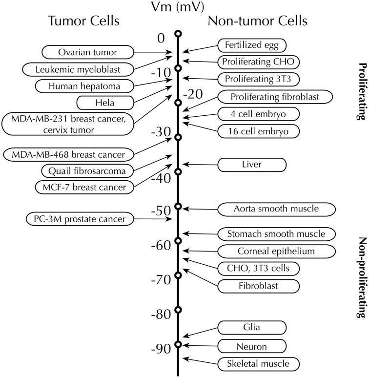
Nothing works without electric potential. That's what makes electrons flow just like water flows from higher potential to lower potential. Ray did not believe in ion pumps but Ray had limited knowledge in cellular electricity. Below from Wikipedia:

Membrane potential


Membrane potential (also transmembrane potential or membrane voltage) is the difference in electric potential between the interior and the exterior of a biological cell. With respect to the exterior of the cell, typical values of membrane potential range from –40 mV to –80 mV.

All animal cells are surrounded by a membrane composed of a lipid bilayer with proteins embedded in it. The membrane serves as both an insulator and a diffusion barrier to the movement of ions. Transmembrane proteins, also known as ion transporter or ion pump proteins, actively push ions across the membrane and establish concentration gradients across the membrane, and ion channels allow ions to move across the membrane down those concentration gradients. Ion pumps and ion channels are electrically equivalent to a set of batteries and resistors inserted in the membrane, and therefore create a voltage between the two sides of the membrane.

Virtually all eukaryotic cells (including cells from animals, plants, and fungi) maintain a non-zero transmembrane potential,[citation needed] usually with a negative voltage in the cell interior as compared to the cell exterior ranging from –40 mV to –80 mV. The membrane potential has two basic functions. First, it allows a cell to function as a battery, providing power to operate a variety of "molecular devices" embedded in the membrane. Second, in electrically excitable cells such as neurons and muscle cells, it is used for transmitting signals between different parts of a cell. Signals are generated by opening or closing of ion channels at one point in the membrane, producing a local change in the membrane potential. This change in the electric field can be quickly affected by either adjacent or more distant ion channels in the membrane. Those ion channels can then open or close as a result of the potential change, reproducing the signal.

In non-excitable cells, and in excitable cells in their baseline states, the membrane potential is held at a relatively stable value, called the resting potential. For neurons, typical values of the resting potential range from –70 to –80 millivolts; that is, the interior of a cell has a negative baseline voltage of a bit less than one-tenth of a volt. The opening and closing of ion channels can induce a departure from the resting potential. This is called a depolarization if the interior voltage becomes less negative (say from –70 mV to –60 mV), or a hyperpolarization if the interior voltage becomes more negative (say from –70 mV to –80 mV). In excitable cells, a sufficiently large depolarization can evoke an action potential, in which the membrane potential changes rapidly and significantly for a short time (on the order of 1 to 100 milliseconds), often reversing its polarity. Action potentials are generated by the activation of certain voltage-gated ion channels.


Ions and the forces driving their motion[edit]

Main articles: Ion, Diffusion, Electrochemical gradient, and Electrophoretic mobility

Electrical signals within biological organisms are, in general, driven by ions.[3] The most important cations for the action potential are sodium (Na+) and potassium (K+).[4] Both of these are monovalent cations that carry a single positive charge. Action potentials can also involve calcium (Ca2+),[5] which is a divalent cation that carries a double positive charge. The chloride anion (Cl−) plays a major role in the action potentials of some algae,[6] but plays a negligible role in the action potentials of most animals.[7]

Ions cross the cell membrane under two influences: diffusion and electric fields. A simple example wherein two solutions—A and B—are separated by a porous barrier illustrates that diffusion will ensure that they will eventually mix into equal solutions. This mixing occurs because of the difference in their concentrations. The region with high concentration will diffuse out toward the region with low concentration. To extend the example, let solution A have 30 sodium ions and 30 chloride ions. Also, let solution B have only 20 sodium ions and 20 chloride ions. Assuming the barrier allows both types of ions to travel through it, then a steady state will be reached whereby both solutions have 25 sodium ions and 25 chloride ions. If, however, the porous barrier is selective to which ions are let through, then diffusion alone will not determine the resulting solution. Returning to the previous example, let's now construct a barrier that is permeable only to sodium ions. Now, only sodium is allowed to diffuse cross the barrier from its higher concentration in solution A to the lower concentration in solution B. This will result in a greater accumulation of sodium ions than chloride ions in solution B and a lesser number of sodium ions than chloride ions in solution A.

This means that there is a net positive charge in solution B from the higher concentration of positively charged sodium ions than negatively charged chloride ions. Likewise, there is a net negative charge in solution A from the greater concentration of negative chloride ions than positive sodium ions. Since opposite charges attract and like charges repel, the ions are now also influenced by electrical fields as well as forces of diffusion. Therefore, positive sodium ions will be less likely to travel to the now-more-positive B solution and remain in the now-more-negative A solution. The point at which the forces of the electric fields completely counteract the force due to diffusion is called the equilibrium potential. At this point, the net flow of the specific ion (in this case sodium) is zero.


Action potential

From Wikipedia, the free encyclopedia


In physiology, an action potential occurs when the membrane potential of a specific axon location rapidly rises and falls:[1] this depolarisation then causes adjacent locations to similarly depolarise. Action potentials occur in several types of animal cells, called excitable cells, which include neurons, muscle cells, endocrine cells, and in some plant cells.

In neurons, action potentials play a central role in cell-to-cell communication by providing for—or, with regard to saltatory conduction, assisting—the propagation of signals along the neuron's axon towards synaptic boutons situated at the ends of an axon; these signals can then connect with other neurons at synapses, or to motor cells or glands. In other types of cells, their main function is to activate intracellular processes. In muscle cells, for example, an action potential is the first step in the chain of events leading to contraction. In beta cells of the pancreas, they provoke release of insulin.[a] Action potentials in neurons are also known as "nerve impulses" or "spikes", and the temporal sequence of action potentials generated by a neuron is called its "spike train". A neuron that emits an action potential, or nerve impulse, is often said to "fire".

Action potentials are generated by special types of voltage-gated ion channels embedded in a cell's plasma membrane. These channels are shut when the membrane potential is near the (negative) resting potential of the cell, but they rapidly begin to open if the membrane increases to a precisely defined threshold voltage, depolarising the transmembrane potential. When the channels open, they allow an inward flow of sodium ions, which changes the electrochemical gradient, which in turn produces a further rise in the membrane potential. This then causes more channels to open, producing a greater electric current across the cell membrane, and so on. The process proceeds explosively until all of the available ion channels are open, resulting in a large upswing in the membrane potential. The rapid influx of sodium ions causes the polarity of the plasma membrane to reverse, and the ion channels then rapidly inactivate. As the sodium channels close, sodium ions can no longer enter the neuron, and then they are actively transported back out of the plasma membrane. Potassium channels are then activated, and there is an outward current of potassium ions, returning the electrochemical gradient to the resting state. After an action potential has occurred, there is a transient negative shift, called the afterhyperpolarization.

In animal cells, there are two primary types of action potentials. One type is generated by voltage-gated sodium channels, the other by voltage-gated calcium channels. Sodium-based action potentials usually last for under one millisecond, but calcium-based action potentials may last for 100 milliseconds or longer.[2] In some types of neurons, slow calcium spikes provide the driving force for a long burst of rapidly emitted sodium spikes. In cardiac muscle cells, on the other hand, an initial fast sodium spike provides a "primer" to provoke the rapid onset of a calcium spike, which then produces muscle contractions.


Overview[edit]


Shape of a typical action potential. The membrane potential remains near a baseline level until at some point in time, it abruptly spikes upward and then rapidly falls.

Nearly all cell membranes in animals, plants and fungi maintain a voltage difference between the exterior and interior of the cell, called the membrane potential. A typical voltage across an animal cell membrane is −70 mV. This means that the interior of the cell has a negative voltage of approximately one-fifteenth of a volt relative to the exterior. In most types of cells, the membrane potential usually stays fairly constant. Some types of cells, however, are electrically active in the sense that their voltages fluctuate over time. In some types of electrically active cells, including neurons and muscle cells, the voltage fluctuations frequently take the form of a rapid upward spike followed by a rapid fall. These up-and-down cycles are known as action potentials. In some types of neurons, the entire up-and-down cycle takes place in a few thousandths of a second. In muscle cells, a typical action potential lasts about a fifth of a second. In some other types of cells, and also in plants, an action potential may last three seconds or more.

The electrical properties of a cell are determined by the structure of the membrane that surrounds it. A cell membrane consists of a lipid bilayer of molecules in which larger protein molecules are embedded. The lipid bilayer is highly resistant to movement of electrically charged ions, so it functions as an insulator. The large membrane-embedded proteins, in contrast, provide channels through which ions can pass across the membrane. Action potentials are driven by channel proteins whose configuration switches between closed and open states as a function of the voltage difference between the interior and exterior of the cell. These voltage-sensitive proteins are known as voltage-gated ion channels.

Process in a typical neuron[edit]


Approximate plot of a typical action potential shows its various phases as the action potential passes a point on a cell membrane. The membrane potential starts out at −70 mV at time zero. A stimulus is applied at time = 1 ms, which raises the membrane potential above −55 mV (the threshold potential). After the stimulus is applied, the membrane potential rapidly rises to a peak potential of +40 mV at time = 2 ms. Just as quickly, the potential then drops and overshoots to −90 mV at time = 3 ms, and finally the resting potential of −70 mV is reestablished at time = 5 ms.

All cells in animal body tissues are electrically polarized – in other words, they maintain a voltage difference across the cell's plasma membrane, known as the membrane potential. This electrical polarization results from a complex interplay between protein structures embedded in the membrane called ion pumps and ion channels. In neurons, the types of ion channels in the membrane usually vary across different parts of the cell, giving the dendrites, axon, and cell body different electrical properties. As a result, some parts of the membrane of a neuron may be excitable (capable of generating action potentials), whereas others are not. Recent studies[citation needed] have shown that the most excitable part of a neuron is the part after the axon hillock (the point where the axon leaves the cell body), which is called the initial segment, but the axon and cell body are also excitable in most cases.

Each excitable patch of membrane has two important levels of membrane potential: the resting potential, which is the value the membrane potential maintains as long as nothing perturbs the cell, and a higher value called the threshold potential. At the axon hillock of a typical neuron, the resting potential is around –70 millivolts (mV) and the threshold potential is around –55 mV. Synaptic inputs to a neuron cause the membrane to depolarize or hyperpolarize; that is, they cause the membrane potential to rise or fall. Action potentials are triggered when enough depolarization accumulates to bring the membrane potential up to threshold. When an action potential is triggered, the membrane potential abruptly shoots upward and then equally abruptly shoots back downward, often ending below the resting level, where it remains for some period of time. The shape of the action potential is stereotyped; this means that the rise and fall usually have approximately the same amplitude and time course for all action potentials in a given cell. (Exceptions are discussed later in the article). In most neurons, the entire process takes place in about a thousandth of a second. Many types of neurons emit action potentials constantly at rates of up to 10–100 per second. However, some types are much quieter, and may go for minutes or longer without emitting any action potentials.

Membrane potential and cancer Membrane potential and cancer progression

Membrane potential (Vm), the voltage across the plasma membrane, arises because of the presence of different ion channels/transporters with specific ion selectivity and permeability. Vm is a key biophysical signal in non-excitable cells, modulating important cellular activities, such as proliferation and differentiation. Therefore, the multiplicities of various ion channels/transporters expressed on different cells are finely tuned in order to regulate the Vm. It is well-established that cancer cells possess distinct bioelectrical properties. Notably, electrophysiological analyses in many cancer cell types have revealed a depolarized Vm that favors cell proliferation. Ion channels/transporters control cell volume and migration, and emerging data also suggest that the level of Vm has functional roles in cancer cell migration. In addition, hyperpolarization is necessary for stem cell differentiation. For example, both osteogenesis and adipogenesis are hindered in human mesenchymal stem cells (hMSCs) under depolarizing conditions. Therefore, in the context of cancer, membrane depolarization might be important for the emergence and maintenance of cancer stem cells (CSCs), giving rise to sustained tumor growth. This review aims to provide a broad understanding of the Vm as a bioelectrical signal in cancer cells by examining several key types of ion channels that contribute to its regulation. The mechanisms by which Vm regulates cancer cell proliferation, migration, and differentiation will be discussed. In the long term, Vm might be a valuable clinical marker for tumor detection with prognostic value, and could even be artificially modified in order to inhibit tumor growth and metastasis.

 
Last edited:
EMF Mitigation - Flush Niacin - Big 5 Minerals

Similar threads

Back
Top Bottom